Art Institute of Chicago
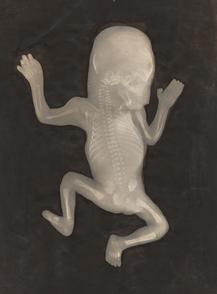
Degree of Ossification of a Five-Month-Old Fetus (Degré d'ossification d'un Foetus de cinq mois)
Léon Tissier
- Date
- 1898
- Medium
- Gelatin silver print with applied india ink or watercolor
- Culture
- France
- Department
- Photography and Media
- Institution
- Art Institute of Chicago
In late 1895, physicist Wilhelm Conrad Röntgen inadvertently discovered a form of radiation that exposed photographic plates; as electromagnetic waves passed through opaque materials, they left behind shadowy images. He termed them X-rays, and news of the discovery spread rapidly. For the scientific community, X-rays expanded medical possibilities, allowing doctors to see interior anatomy without surgery. For the general public, these pictures spectacularly revealed places normally invisible to the human eye and became objects of enduring fascination. This X-ray appeared on the cover of Les Rayons X , a journal of radiology, on March 5, 1898, just two years after Röntgen's discovery. The accompanying article claimed that X-rays were "interesting and useful for the study of fetal development at all stages of pregnancy," describing in wonderment what can be seen clearly in the image (larger bones and the spine) and what cannot (connective tissue and cartilage).
The authoritative record is held by Art Institute of Chicago. LinkedCulture surfaces this object and its connections; it does not alter institutional metadata.
Linked open data
Authority identifiers that link this record into the wider web of cultural data — stable references you can follow to the source.
- Object type
- AAT300046300
Related across collections
Semantically similar works from Art Institute of Chicago and other institutions.
![[Diogenes without Sun, 17' Exposure]](https://media.getty.edu/iiif/image/fe9d2cd2-3a22-4bc7-aed3-2e8eff50b362/full/808,/0/default.jpg)
[Diogenes without Sun, 17' Exposure]
Getty Museum
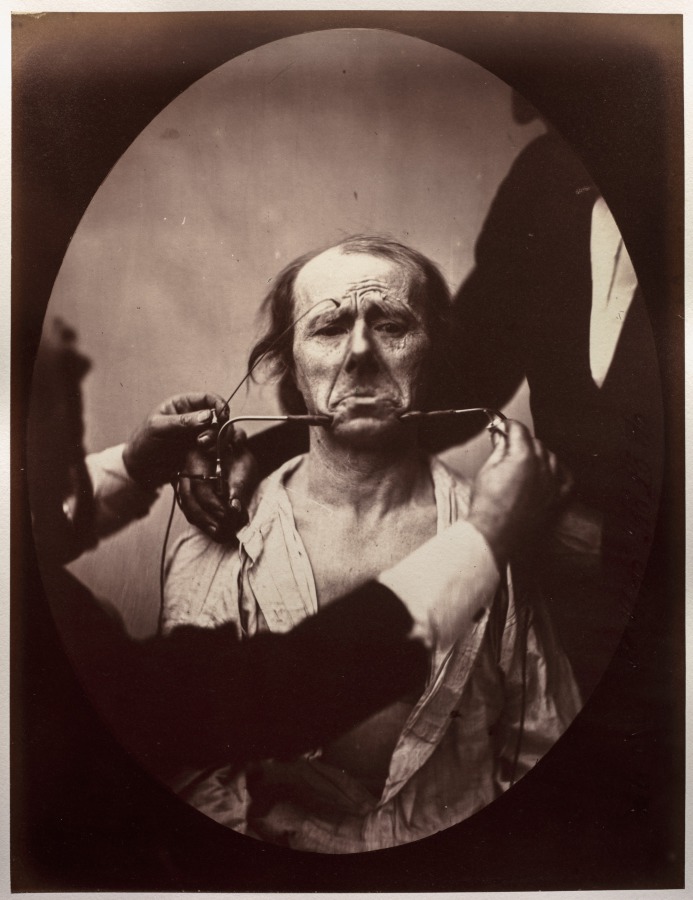
Figure 45: Contraction électrique forte des triangulaires des lèvres et des sourciliers: douleur et déspoir
Cleveland Museum of Art

Portrait of a Father and Smiling Child
Getty Museum
![[Postmortem Portrait of a Baby]](https://media.getty.edu/iiif/image/45b8eab1-57cc-428e-9402-0b667052de00/full/808,/0/default.jpg)
[Postmortem Portrait of a Baby]
Getty Museum
Mother and Child
Cleveland Museum of Art
![[Study of a white foal]](https://media.getty.edu/iiif/image/344b361a-91c5-49d2-9d71-8ae8667bad40/full/808,/0/default.jpg)
[Study of a white foal]
Getty Museum
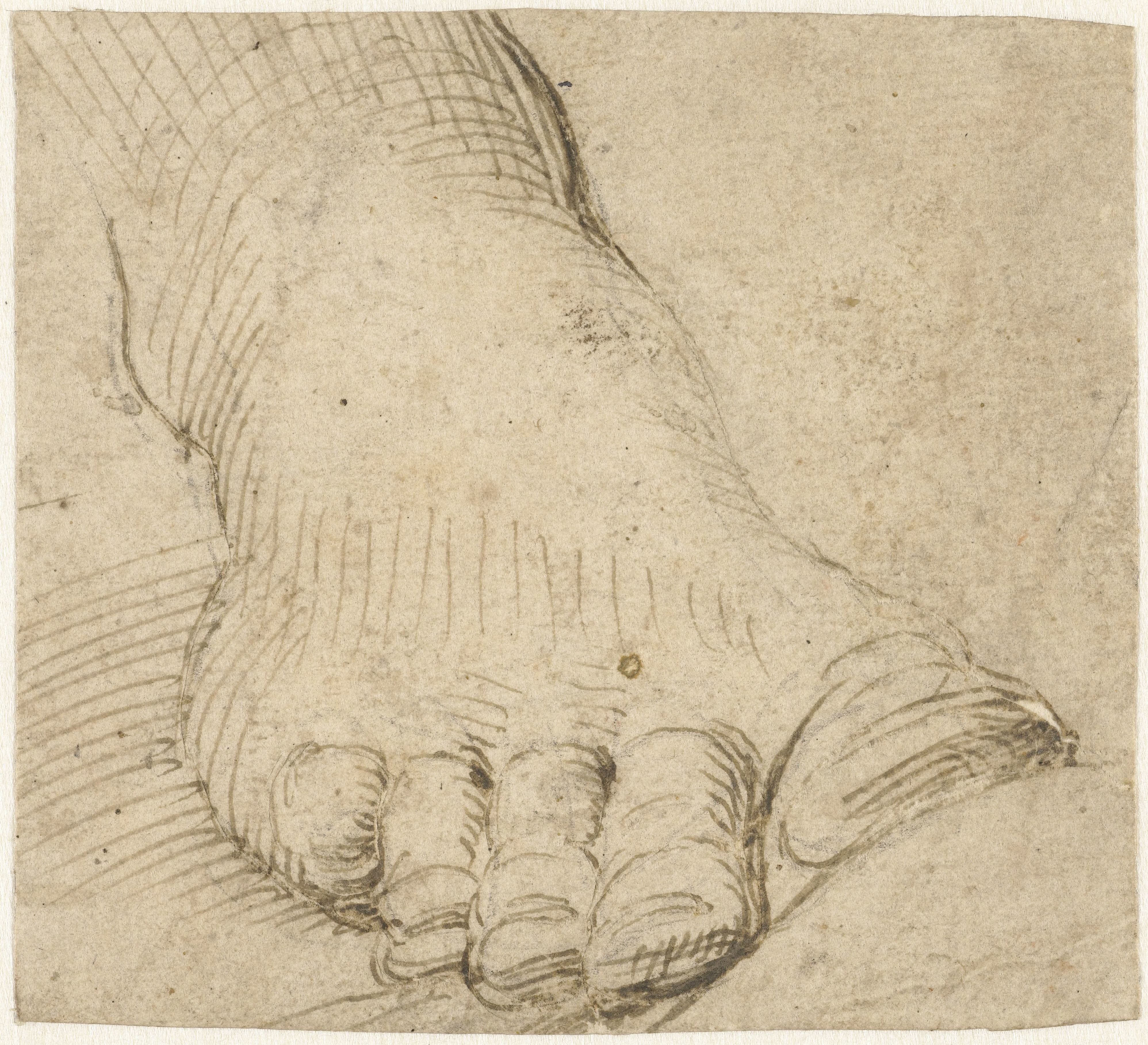
Studie van een rechtervoet, van voren
Rijksmuseum

Young Bourgeois Mother
Getty Museum
Untitled (George Coit)
Art Institute of Chicago
![[Postmortem Portrait of an Infant]](https://media.getty.edu/iiif/image/6fb6deb6-9445-461d-8a5c-ff19aa5b2a13/full/808,/0/default.jpg)
[Postmortem Portrait of an Infant]
Getty Museum

In the Snow
Minneapolis Institute of Art

Bust of Venus, November 26, 1840
Getty Museum